Liposome formation tutorial

DSCF liposomes

Liposomal encapsulation serves as the basis for the engineering of biomimetic and novel synthetic cells. Liposomes are normally formed using such methods as thin film rehydration (TFH), density mediated reverse emulsion encapsulation (REE), or one of many microfluidics-based approaches. While several microfluidics-based methods exist, capable of efficiently forming unilamellar liposomes, with uniform size and acceptable encapsulation rates, the main limitation associated with microfluidics is that trace amounts of carrier organic solvent remain present in the resultant membrane. A popular method bypasses the problem of residual solvent presence by first, evaporating all carrier solvent and thereby creating a thin lipid film used to prepare liposomes through subsequent methods. However, most protocols which utilize this methodology of thin film preparation are non-microfluidic protocols and thus do not produce uniform unilamellar liposomes. 

DSCF, which stands for Droplet‐Shooting Centrifugal Formation, is a derivative liposome formation method that utilizes solvent-less lipid thin films to prepare highly uniform lipid bilayer membranes using a 3D printable microfluidics-system. In this way, DSCF methods avoid the solvent issues associated with microfuidics while producing liposomes with a high level of repeatability, similarly to microfluidics.

Various protocols for the preparation of DSCF liposomes exist whereby lipid-bilayer membranes are formed when lumen droplets pass through a lipids-in-oil solution and thence into an aqueous solution via centrifugal force. Utilizing this general DSCF mechanism it is possible to assemble highly uniform liposomes quickly and easily with tunable lumen and membrane chemistries. 

adamala lab liposome formation

Device

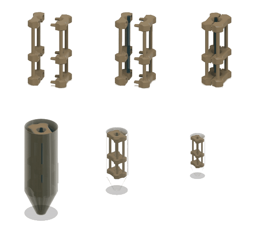
DSCF liposome formation is based, in part, on DSSF (droplet shooting size filtration) and similarly utilizes a micro capillary collet that holds a glass capillary within a micro centrifuge tube. However the DSCF device is composed of a standard micro centrifuge tube, a commercially available pre-pulled microcapillary, and a 3D printable collet.
As compared to DSSF the 3D printed DSCF device is easier to assemble by hand and avoids challenges associated with capillary-collet assembly.

The DSCF device may be 3D printed using an SLA printer or an online 3D printing service. Additionally, an injection molding form has been manufactured by the Adamala Lab for the production of micro capillary collets in Polyetherimide (PEI), an amorphous chemically resistant plastic. 

Get the device from us

Due to supply chain issues, we have to discontinue our injection molded devices program.

We are working on finding a new supplier of affordable parts, we will resume shipping whenever possible.

3D print your own device

You can download files to 3D print your own device.

Download .step file.

Protocol

Follow the general protocol below. For more details and example data, see 
https://link.springer.com/protocol/10.1007/978-1-0716-1998-8_14

1 Materials

Prepare all aqueous solutions using ultrapure water (prepared by purifying deionized water, to attain a sensitivity of 18 MΩ-cm at 25 °C). For non-polar preparations, use glass pipettes, adhere to proper safety precautions regarding volatile agents, and follow all waste disposal regulations when disposing of waste materials.

1.1 3D-Printed Microfluidics Device

1. 3D-printed microcapillary holder: SLA printed microcapillary holder, printed in Accura 60 material. Printed in high resolution (XY plane: +/- 0.005” for the first inch, plus +/- 0.002” for every inch thereafter. Z plane: +/- 0.010” for the first inch, plus +/- 0.002” for every inch thereafter) (see NOTE 1).
2. Glass microcapillary: 1 mm OD microcapillary with 10 um pulled tip.
3. Glass microcapillary scoring file: Blade shaped medium grit ruby degussit abrasive file.
4. Conical centrifuge tube: 1.7 mL conical microcentrifuge tube with 2 cm throat length and 9 mm ID.


1.2 Lipid-Oil Solution

1. Lipid-Oil Solution use in this demonstration was: 4.925 mM POPC, 4.925 mM DOPC, 0.15 mM fluorescently labeled PE (NBD-PE, N-(7-Nitrobenz-2-Oxa-1,3-Diazol-4-yl)-1,2-Dihexadecanoyl-sn-Glycero-3-Phosphoethanolamine).
2. Prepare a 1,2-dioleoyl-sn-glycero-3-phosphocholine (DOPC) and 1-palmitoyl-2-oleoyl-glycero-3-phosphocholine (POPC) stock solutions by adding a non-specific amount of each powdered lipid species to separate massed glass vials.
3. Mass the vials to determine the amount of each lipid present within their respective vials.
4. Dissolve the powdered lipids within the vial in 5 mL of CHCl3 and recorded the measured concentrations of each solution using the observed mass of each lipid.
5. Use these solutions of known concentrations to prepare a singular 1:1 9.85 mM DOPC/POPC solution; however, do not adjust this solution to its final volume yet.
6. Add 0.15 mM of the desired fluorescently labeled lipid and subsequently bring the solution up to its final volume. At this point one should be left with a 10 mM 1:1 DOPC/POPC solution doped to 1.5 mol% with a fluorescently labeled lipid.
7. Aliquot 20 uL (for a 1 mM lipid-oil solution) or 10 uL (for a 0.5 mM lipid-oil solution) of this solution into brown HPLC glass vials.
8. Leave to desiccate inside a fume hood for minimum 24 hours, or desiccate under gas flow. (see NOTE 2 and NOTE 3).


1.3 Biomimetic Lumen Chemistries

Example RNA aptamer Broccoli transcription
1. The reaction was prepared as previously described, with the final concentration of all reagents: 4 mM each NTP, 40 mM tris pH 7.9, 42mM MgCl2, 100 mM KCl, 2 mM spermidine, 1 mM DTT, 2 μL 1 mM DFHBI-1T Broccoli aptamer ligand, 2 μL 5 μM Oligonucleotide Broccoli DNA template duplex, 2 μL 10X T7 RNA polymerase, 2 μL 10X Inorganic pyrophosphatase.
2. The Broccoli template used in this work was, the sense strand: d(TAA TAC GAC TCA CTA TAG GAG ACG GTC GGG TCC AGA TAT TCG TAT CTG TCG AGT AGA GTG TGG GCT C).

Example TX;TL of Green Fluorescent Protein (GFP).
1. The reaction was prepared as described before, using TxTl lysate of E. coli Rosetta DE3 strain and sonication protocol.
2. The final concentration in the reaction mixture was 500 mM HEPES pH 8, 15 mM ATP and GTP, 9 mM CTP and UTP, 2 mg/mL of E. coli tRNA mixture, 0.68 mM folinic acid, 3.3 mM nicotinamide adenine dinucleotide (NAD), 2.6 mM coenzyme-A (CoA), 15 mM spermidine, 40 mM sodium oxalate, 7.5 mM cAMP, 300 mM 3-PGA, 12mM Mg-glutamate, 140 mM K-glutamate, 1 mM DTT, 2 mM each of 20 amino acids, 10 nM GFP plasmid, RNAse inhibitor Murine 40U/ul 1x, 1 uM T7 RNA polymerase, cell free lysate 0.33x total reaction volume.

1.4 Liposome Preparation Buffer

1. To make liposome preparation buffer: mix 100mM HEPES, 900mM Glucose, pH 7.5.
2. Add about 100 mL water to a 1 L graduated cylinder or glass beaker.
3. Weigh 23.83 g HEPES and transfer to cylinder. Weigh 162.14 g Glucose and transfer to cylinder.
4. Add water to a volume of 900 mL.
5. Mix and adjust pH with HCl and NaOH respectively. Make up to 1 L with water.
6. Filter sterilize the buffer. Store at 4 °C.
2 Methods

Carry out all procedures at room temperature unless otherwise specified.


2.1 Preparation of Lipid-Oil Solution

Add 200 uL of mineral oil to desiccated lipid films. (see NOTE 5).
Sonicate the mineral the lipid-oil solution to expedite solvation. Sonicate for two periods of 30 minutes with intermediate high intensity vortexing before and after each period of sonication.

2.2 Assembly of 3D-Printable Microfluidics Device
1. Test fit male and female subunits of 3D printed microcapillary collet chassis and insure proper subunit fit-up. (see NOTE 5).
2. When using 1.7 mL microcentrifuge tubes with a 2 cm throat length, score microcapillaries and break to a length of approximately 24 mm using a ruby degussit abrasive file. (see NOTE 6).
3. Carefully, as to not damage the delicate pulled tip of the microcapillary, seat the glass capillary in the central channel of the male subunit using a forceps to manipulate the microcapillary with the subunit resting on an elevated flat surface. Once the capillary is in place within the central channel, assemble the two subunits together by registering the pins of the male subunits to the ports of the female subunit. (see NOTE 7).
4. Aliquot 30 uL of liposome preparation buffer into the bottom of a 1.7 mL microcentrifuge tube making sure that the volume ends up in the posterior terminus of the conic. (see NOTE 8).
5. Layer 100 uL of the Lipid-Oil solution on top of the 30 uL of liposome preparation buffer by aliquoting the 100 uL while carefully swirling the tip of the pipette circularly around the internal circumference of the microcentrifuge tube. (see NOTE 9).
6. Cap and let equilibrate for 30 minutes at STP. (see NOTE 10).
7. Carefully insert the 3D printed microcapillary collet into the microcentrifuge tube.


3 Liposome Preparation
1. Taking advantage of capillary action, use a pipette to carefully fill the seated microcapillary, within the 3D printed microfluidics device, with the desired lumen chemistry (See section 2.3).
2. Cap assembly and subsequently centrifuge at 1,600 RCF for 3 minutes.
3. After centrifugation, remove the 3D printed collet and microcapillary from the microcentrifuge tube using serrated forceps.
4. Aspirate off 29 uL of the aqueous bottom layer.
5. Carry out data collection, using method specific to the lumen chemistry.

4 Notes

1. 3D-printed parts can be ordered from Xometry (https://www.xometry.com) or any other 3D printing service. STL and STEP files are attached as supplementary information with this paper.

2. Preparing lipid stock solutions can aid in the preparation of lipid solutions as it can be difficult to accurately measure and handle lipid powders due to their unguent physical properties. Additionally, working with volumes of CHCl3 in volumes of 5 mL or more can decrease the concentration skew associated with evaporation. Moreover, a (2:1) mixture of CHCl3 and CH3OH may be used to prepare non-aqueous lipid solutions.

3. Glass syringes (e.g. Hamilton 1700 Series Syringes) should be used for the handling of lipid solutions dissolved in nonpolar solvents. Syringes should be appropriately cleaned and sterilized in order to prepare accurate solutions.


4. Our experimentation has shown that mixed weight mineral oil seems to be ideal for liposome preparation using this method (i.e. not light or heavy mineral oil).


5. Seating and unseating the male and female subunits can help mold the pin geometry and facilitate a proper fit-up.


6. Using your finger as a stop to gently glide the file around the circumference of the microcapillary can help to create a less jagged edge; doing this not only creates a safer edge but is itself also a safer process as it results in less microcapillary fragmentation.


7. The seating of a glass microcapillary can somewhat retract in the superior direction prior to the mating of the two subunits as once the subunits are combined the capillary can be fulling seated by applying light axial pressure downwards from the superior terminus of the microcapillary.


8. A benchtop centrifuge may be used to ensure that the droplet of buffer is fully in the bottom of the microcentrifuge tube.


9. Gel loading tips can greatly aid in this step as their flexibility makes the swirling motion easier.


10. Equilibration is necessary for the oil solution to fully settle down the side of the microcentrifuge tube.


11. During encapsulation, the TxTl solution is diluted (accounting for losses from incomplete encapsulation). Also, the lumen of liposomes only comprises of fraction of the volume of the whole sample. Therefore, equal volume comparison for encapsulated and non-encapsulated TxTl solutions will not provide reliable indication of performance of the encapsulated solution. We provide 1x (=liposome sample volume) and 0.25x (=quarter of liposome sample volume) unencapsulated controls.


12. The fluorescein solution should remain stable over the time of the experiment. Any change in fluorescence from the marker solution indicates evaporation from incompletely sealed plate, or some other technical problem.

The team

All work was done in the Adamala lab.

The device was designed and made by Orion Venero.

The protocol was validated and refined with the help of Wakana Sato, Joseph Heili and Christopher Deich. Kate Adamala helped by not getting in the way.

Funding

This work was supported by the National Science Foundation award 1844313, RoL: RAISE: DESYN-C3: Engineering multi-compartmentalised synthetic minimal cells, by the National Aeronautics and Space Administration grant 80NSSC18K1139, Center for the Origin of Life - Translation, Evolution And Mutualism; and by the John Templeton Foundation grant 61184, Exploring the Informational Transitions Bridging Inorganic Chemistry and Minimal Life.

Citation

If you use this protocol and/or the device, please cite:

Venero O.M., Sato W., Heili J.M., Deich C., Adamala K.P. (2022) Liposome Preparation by 3D-Printed Microcapillary-Based Apparatus. Methods in Molecular Biology, vol 2433. Humana, New York, NY. https://doi.org/10.1007/978-1-0716-1998-8_14

https://link.springer.com/protocol/10.1007/978-1-0716-1998-8_14